The anatomical sources on which iridology relies are three: the iris surface, the pupil and the sclera. Each of these structures provides information which integrates patient’s evaluation.
Main iridology markings
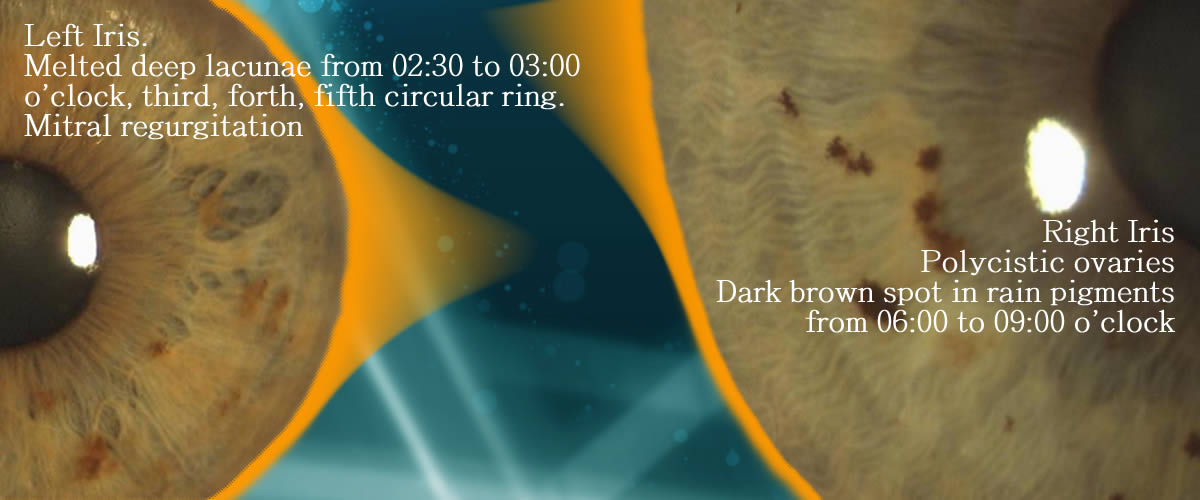
Lacunae. Oval or lozange shaped area without fibers or with very sparse one.
Bright fibers. Lightening of texture.
Transversal fibers. Fibers not following the radial pattern of texture.
Radial furrows, fissured arcs. Spasm and contractions.
Pigments. Iris spots that can take various shapes and colors.
Pupillary flattening. Straightened segment of pupillary edge with loss of roundeness.
THE IRIS SURFACE
Fibers
Fibers are the surface radial structure of iris stroma. Fibers are in a normal condition when they spread radially and are tight together with no overlapping. According to their diameter, they are called velvet, silk, linen or hemp-like fibers. An enlarged diameter is associated with an increased thinning of fiber texture.
Enlarged white fibers. White suffusions. The increase in fiber diameter and a fiber color turning to white or bright white is a sign of inflammation, either an ongoing or a recent process.White impregnations in adjacent areas are a worsening sign.
Fiber thinning. Fiber thinning is accompanied by a darkening in the thinned-out areas between fibers due to lower layers of the stroma becoming visible.
Transversal fibers. These are fibers showing a larger diameter than that of stroma fibers and with a transversal rather than a radial direction. Transversal fibers can be simple, branched, grouped in bundles or circumscribing small sectors of the iris like an arc.
Vascularized transversal fibers. Transversal fibers with a visible blood vessel.
Tulip Fibers. Change of the basic texture by creating a darker area which acquires various shapes (tulip marking, asparagus marking).
Abnormal fibers. They circumscribe arc or angle-shaped areas.
Silver fibers. Thin white fibers which are much paler than stroma fibers.
Neuronal net. Widespread thick networks of thinner fibers which lay upon stroma fibers.
Kidney bridges. Fibers circumscribing wide arcs which are concave towards the ciliary body edge in the lower sector. They reveal kidney disorders or a mineral and water imbalance.
Lightening. Areas showing a lighter color and visible white fibers.
Koch’s bridge. Frayed abnormal fiber detaching from the collarette and inserting in other iris areas like a bridge.
Lacunae.
Shape of lacunae. Several studies have associated variations in the morphology of lacunae with diseases. Deviations from the standard oval shape are to be interpreted as warning signs triggering the attention threshold although they do not provide sure disease diagnoses. In-depthinformation on the subject can be found inA. A. Santorelli (Semeiotica dell’iride” ASSIRI, 2006), F. Minisini, S. Pizzini (“Il rimedio dall’iride”, M.I.R. ed. 2008), J. L. Berdonces (“Trattato di Iridologia”, Red ed. 2003, El gran libro de la iridologia).
Following is a concise re-elaboration of the above sources:
- Torpedo or cigar lacuna. It pushes against or breaks into the wreath. Glandular tumors. If located in the medial-lower part of the lateral sectors, it can be a sign of breast cancer. Perifocal signs (lightening of iris color, pigments, transversal fibers) point to a worsening process.
- Daisy petal lacuna. Lymphatic system pathologies.
- Curved beak lacuna. Lacuna with a curved end inserting into the collarette. Predisposition to degenerative pathologies in the genital area.
- Asparagus lacuna. It can also manifest with rarefaction of fibers. Degenerative pathologies in the urogenital area.
- Iodine lacuna. Its base is located against the wreath and its end is turned outwards. Thyroid diseases.
- Grape lacuna. Cluster of small dark lacunae. Goiter, lymphatic glands diseases.
- Solitary lacuna in the thyroid zone. It is called thyroxin lacuna.
- Half lacuna. Functional problems in the corresponding organ.
- Leaf lacunae. They are generally found in the lower part of the iris and are associated with endocrine disorders. If they insert into the autonomic nerve ring, they relate to a worsening condition.
- Bean lacuna. It is located agains the autonomic nerve ring. Endocrine dysfunctions (thymus), psychosis.
- Twin lacunae. Pair of lacunae located at 6 o’clock in the adrenal zone. Adrenal defects.
- Stairstep or rooftile lacuna. It originates from the autonomic nerve ring and extends towards the ciliary border. Predisposition to degenerative pathologies.
- Double lacuna. It is divided into two parts by a thick margin. Anemia. If found in the heart zone, it is related to cardiovascular pathologies.
- Negative lacuna. A lacuna which is sunken as a consequence of the raising of the surrounding iris surface. Metabolic disorders: gout, diabetes, organ hypertrophy, hepatomegaly, splenomegaly.
Cramps and contractions
- Radial or circular furrows embedded in the iris texture.
- Circular furrows or rings are concentric to the pupil and are called contraction rings, tetanic rings, spasm rings or cramp rings.
- Radial furrows are also called radii solaris or iris rays.
- These signs characterize the spasmodic iris type.
Contraction rings (tetanic rings)
- Tetanic rings. Circular concentric furrows located in the ciliary body. They indicate a predisposition to spasms and contractions and relate to heart neurosis, anxiety somatization, panic attacks, sleep disorders. They cross wide sectors of the iris; the starting and ending point of the ring indicate the main organs affected. The more the rings are, the stronger the predisposition to spasmophilia is. Rings located close to each other or even covering the whole iris circonference are considered as a worsening sign.
- Concentric rings. More than one ring in the same area indicate a predisposition to spasmophilia in the corresponding organs.
- Intersecting rings?. They indicate spasms in the intersecting point.
- Ring inside the crown. It is called tachyphagic ring. Gastric and gastro-intestinal spasms.
- Oppression rings. More than one contraction ring with a vertical orientation enclosing the lateral sectors (temporal and nasal). Heart and respiratory oppression, asthma, bronchial spasm.
Radii solaris
- Radial contraction furrow starting from the pupillary border or from the autonomic nerve wreath.
- The radii solaris starting from the pupil are called radii major while those starting from the wreath are called radii minor.
- Radii major reveal a greater predisposition to spasmophilia while radii minor relate to digestive spasmophilia.
- Radii solaris indicate a predisposition to headache, physical or mental breakdown, decrease of attention and memory.
- The radii major starting from the brain zone of the pupil and reaching the frontal sector indicate a predisposition to digestive headache.
- The radii minor starting from the wreath in the frontal sector associated with contraction arcs reveal a predisposition to endocrine disorders (hypophysis, epiphysis).
Spots, pigments, chromatic impregnations
Spots, pigments and chromatic impregnations are small to large areas showing alterations in the basic iris color. They can appear in different shapes, colors, dimensions. For a better understanding of these important iris signs, it is worth to remind the main classification of the iris according to its basic color: light-colored iris, lymphatic constitution: dark-colored iris, hematogenic constitution; mixed color, biliary constitution. Spots, pigments and chromatic impregnations extend over the constitutional colors.
Classification of pigments
Pigments can be classified into five main categories: urosein, rufin, porphyrin, melanin, gastrin. In addition, other pigments can be identified which characterize the organs they are related to. Also, various basic colours involving the whole iris area can be related to single organs or apparatuses.
Uroseine. Light yellow pigment impregnating wide iris areas through which the iris texture remains visible. It is a result of oxidized carotenoids accumulation. It is related to kidney overload and depurative urinary suffering (kidney stone, gout, kidney failure). A dirt yellow color or yellow tophi point to a predisposition to allergies.
Rufin. Reddish or red brick pigments. Rufin is a derivative of fat transformation (isoprene oxidation).It is a sign of predisposition to liver and pancreas disorders, predominance of oxidation processes, surplus of free radicals, tissue intoxication.
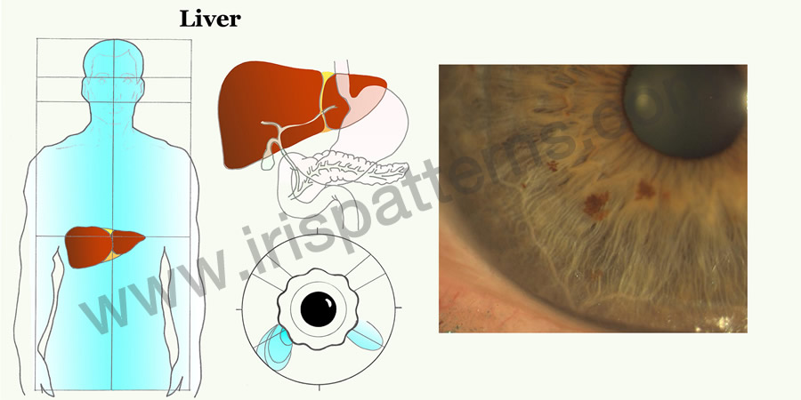
Porphirine. Dark brown pigments deriving from pyrrole, a product resulting from hemoglobin degradation. They indicate liver disorders and can appear in all iris sectors. The presence of porphyrin pigments is related to a bad conversion of hemoglobin. Hazelnut pigments turning brown are interpreted as a worsening sign.
Gastrin. Salmon pink or reddish-orange pigments appearing inside the collarette in the shape of a ribbon or losange. They are related to stomach conditions.
Melanin. Brown or black pigments deriving from the transformation of the tiroxine and phenylalanine amino acids. They are similar to moles. Solitary pigments relate to the organ reflected in the sector in which they appear. They indicate predisposition to degenerative disorders.
Organ-related pigments. Two well-characterized small pigments located respectively in the gastric ring or collarette and in the visceral sector are specifically related to stomach and uterus. A salmon pigment in the shape of a ribbon is typical of stomach disorders while a pink pigment points to minor disorders of the uterus
Colors and organs. Iris color can be associated with a suffering organ: brown is related to liver, orange to pancreas, yellow to kidney, dark brown or black to mucosae and glands.
Location, number, color.
A solitary pigment appearing on a uniform iris color has more significance than the presence of several pigments. The location of the solitary pigment can help identify the suffering organ. When several pigments are distributed in more than one area, it is the pigment color that can help detect weak points.
PUPIL
Normal pupils are alike, uniformly circular and located at the center of the iris. Any deviation from these features reveal weakness or imbalance.
Pupil alterations can be caused by organ distress or spinal column suffering.
The presence of adjacent collarette and ciliary body alterations can provide further insight.
Size alterations. The difference in pupil size is called anisocoria and is a sign of cerebral suffering. The most damaged brain hemisphere is the one corresponding to the eye with a dilated pupil (mydriatic pupil). In case of fixed mydriasis of one or both pupils, there is a high probability of severe acute brain damage (spasm, ischemia, hemorrhage, cerebral trombosis).
Pupil constriction (miosis) can result from narcotics overdose or exposure to toxic substances (insecticides). In acute miosis caused by severe intoxication, extremely constricted pupils are also called “pinpoint” pupils.
Unilateral pupil constriction or unilateral miosis is generally caused by a painful response to organ suffering: kidney cramps, liver cramps, pancreatitis, appendicitis, angina pectoris, optical nerve paralysis.
The Bernard-Horner’s syndrome, that involves only one eye (miosis, droopy eyelids, reduction in eye size), is an example of unilateral miosis with damages limited to the eye.
A rapid and rhytmic sequence of pupillary dilation and constriction unrelated to illumination is called hippus. It is a sign of high nervous reactivity, sensitivity, predisposition to energy exhaustion, use of stimulating substances. The desynchronization between the eyes is a worsening sign.
Pupillary alterations providing useful iridological information are the loss of roundness (ovalization, flattening), changes in pupil position (pupil decentration) and changes in the inner pupillary border (irregularities):
- ovalization
- flattening
- decentration
- inner pupillary border irregularities
Ovalization
Ovalizations consist in loss of roundness of the pupils which tend to elongate and flatten. Pupil axis can be symmetrical and parallel or lean in opposite directions (diverging ovalization). Ovalization can involve one eye only. They suggest nervous irritability or organ distress.
Vertical symmetrical ovalization can be related to cardiovascular or thyroid conditions whereas horizontal symmetrical ovalizations can suggest neurological diseases.
Symmetrical ovalizations inclined to the right or to the left can indicate weakness in the brain area corresponding to their inclination.
Diverging inclinations can delimit wide areas with their axes where possible organ distress can be located.
In case of ovalization of one pupil, the pupil’s axis indicates the upper or lower suffering areas.
Flattening
Flattenings are related to suffering of the spinal column: frontal flattening / cervical column; lateral flattening / dorsal column; ventral flattening / lumbosacral column.
In case the flattening is located in the same “time slot” as accessory signs in the collarette or in the ciliary body (pigments, lacunae, crypts, tophi etc.), defects or suffering of organs projected in that area must be considered.
Pupil decentration
The pupil can lose its central position. Delocalization indicates a moving away from a suffering area. For example, a right pupil shifting toward 2 o’clock is moving away from the liver and gallbladder area (7 o’clock). This might suggest liver and gallbladder distress. Also, a left pupil moving toward the nasal sector should be related to possible heart or left lung suffering.
Bridge.
It is a fiber in the collarette which crosses the pupil and ends in another part of the collarette or iris. It reveals an unstable nervous system.
SCLERA
Sclera pigments are also studied by official medicine. Some colorations have a clinical and other an iridological significance.
Official medicine:
- Yellow or yellowish sclera: cholestasis, liver diseases.
- Reddened sclera: conjunctivitis, congestion, hypertension.
Iridology:
- Light blue or bluish sclera: chronic bone diseases, rheumatic disease, arthrosis, osteoporosis.
- Sclera with brown spots: liver overload.
- Prominent whitish layer similar to mucus: stomach disorders.
More frequently, sclera spots are light brown impregnations that are interpreted as predispositions to liver disorders.
Other common markings are lipid deposits that can calcify due to inflammatory processes. Deposits are often vascularized by a web of perpendicular vessels which are a sign of inflammatory reactivity.
The features of sclera vessel can provide useful information.
Clearly visible solitary vessels pointing toward the iris edge are called pointers. They are considered as indicative of a suffering iris sector. A similar significance is attributed to tangential vessels andfork vessels which, once they have reached the area adjacent to the outer iris border, run perpendicularly to the border touching or delimiting iris areas which are considered as suffering.
Other vessels with abnormal features like dilatations, meanderings, spams, diffusemicro aneurysms can suggest a predisposition to circulatory or coagulation diseases.
Some of these are attributed with specific names: Meander vessel, diving vessel, gun-barrel vessel, pressure-arc vessel, glomerular vessel, arachnoid vessel, ectasia vessel, Meander vessel delimited by two vessels of a smaller diameter. Each morphology is related to a pathological predisposition which, in any case, must be investigated using the diagnostic procedures of official medicine.
Gun-barrel vessel: two parallel vessels with different diameters. They are signs of hemorroids, lower limbs varicose veins and, in case of cirrhosis, portal hypertension and stomach varicose veins.
Meander vessel: large winding vessel delimited by two vessels of a smaller diameter. It suggests lower limbs varicose veins.
Pressure-arc vessel: arched vessel pointing toward the iris indicating a pathology in the iris sector involved.
Ectasia vessel: dilated vessel related to blood stagnation caused by heart conditions, vascular disease, portal stasis.
Spindle-shaped vessel: vessel diameter narrowing progressively.It is indicative of vascular spasm caused by angina pectoris, Reynaud’s syndrome, peripheral vascular diseases.
Porcelain vessel: milk-white, translucent vessel related to dyslipidemia, arteriosclerosis, metabolic syndrome.
Glomerular vessel: ball of capillars reminding of a renal glomerulus suggesting kidney diseases.
Aracnoid vessel: vessel reminding of a spider related to liver-gallbladder diseases and hydroelectrolytic imbalance.
Fork vessel: vessel delimitating suffering areas.
Microaneurysms: capillar fragility, diabetes, coagulation defects, vascular diseases.
Sclera capillaries are normally not visible or barely visible. They appear in case of inflammations orirritativepatologies of nearby organ areas.
The presence of abnormal sclera capillaries (dilatation, spasm, Meander vessels, thick net of capillaries, microaneurysms) can be a sign of vessel or heart diseases or responses to ongoing inflammations.
In addition, the sclera can manifest pigmented toxic metabolites and gather lipid deposits and calcifications in case of altered fat metabolism (hypercholesterolemia, hypertriglyceridemia).
SHARED SIGNS BETWEEN SCLERA AND IRIS
Lunula.
Milk-white deposit around 3 o’clock or 9 o’clock. It suggests predisposition to arteriosclerosis or, if its located in the left temporal sector, predisposition to heart disorders like arrhythmia and tachycardia. It is typical of the cholesterinic diathesis.
Calcific lunula.
It suggests predisposition to arteriosclerosis, high parasympathetic activity with spasmophilia, tendon calcifications, Dupuitren’s syndrome, Peironie’s disease.
Cholesterinic ring/gerontoxon, arcus senilis, sodium ring.
Opaque milk-white ring covering the peripheral area of the iris in the frontal or ventral zone or in a circular band. In its first stage, it is transparent and called sodium ring. It reveals senescence and a predisposition to arteriosclerosis.
Pterygium.
Swelling of the cornea also covering portions of the iris. It can be associated with functional disorders in the organs represented in the iris areas involved. It is sometimes related to brachial plexus disorders.


