IRIS RECOGNITION AND IRIDOLOGY
Iris analysis is used in two main fields: Iris recognition and Iridology.
Iris recognition is a biometric automated method used in the field of privacy and security. Like a snowflake, the iris is unique: a distinctive pattern that forms randomly in uterus in a process called chaotic morphogenesis. It’s estimated that the chance of two iris (irises) being identical is 1 in 1078.
The Iris is the only internal organ visible from outside the body. This allows for a non-intrusive method of capturing an image from a distance of 7,5-15 centimeters away (3 to 10 inches).
The recognition process analyzes the iris patterns that are visible between the pupil and sclera (white of the eye) and converts them into digital template. The iris’ biometric recognition system captures over 240 degrees of unique characteristics in formulating its algorithmic template. The unique characteristics include rings, furrows and freckles within the iris. Iris recognition uses video infrared images, the results are monochromatic images.
Iridology is a complementary medicine technique that analyze the colored portion of the eyes (irises). The irises’ morphology is a good window on persons’ constitution. Iridology and Iris Recognition methods use the same font for different purposes: Iridology for healthcare, Iris Recognition for security. The patterns looked for and analyzed by the tow techniques are the same, but the instruments utilized are different. Lenses and optical instruments (iridoschopes) that give visible spectrum images, for iridological practice. Biometric infrared scanners that give monochromatic images, for iris recognition.
Iridology studies the colored portion of the eye named iris. The iris is a highly innervated organ which is stimulated both by the external environment and as well as by the body. The structure of the iris mirrors the individual constitution; illnesses, harmful habits and aging can alter it. The iris analysis completes medical practice by supplying data on constitution, nervous response, damages caused by aging, illnesses and familiarity. Iridology is a discipline that enriches traditional investigations. It collects signs. It does not provide diagnosis.
IRIS MAPS
The connections and interactions between internal organs and body’s surface are utilized in several fields of health care: Traditional Chinese Acupuncture, Reflexology, Kinesiology …
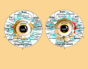
The Hungarian physician I.V. Peczely (1826-1911) realized that this approach could also be applied to the iris surface. In 1871 Peczely started to divulge his discoveries in conferences and publications. In 1880 he published his map of human internal organ projections in the iris (“Entdeckungen auf dem Gebiete der Natur und Heilkunde”, Budapest, 1880). The first physician who recognized the scientific reliability of his discoveries was Emil Schlegel who published, in 1886, a re-elaboration of Peczeley’s original map thus acknowledging his fundamental contributions (“Die Augendiagnose der Dr. Ignacz Von Peczely”, Tubingen, 1886). Peczely and Schlegel are considered as the founders of modern iridology. The version of their maps and diagrams provided in this e-book is a re-elaboration and translation of different sources which aims at respecting the original ones as much as possible.
The main problem with iridological maps is the available space. Unlike acupuncture points, which are distributed all over the body and organized along well separated meridians, the projections of organs in the irises are concentrated in a very limited space. In addition, their graphic representation is one-dimensional while the organs are tri-dimensional and close to each other. As a consequence, different organs necessarily share the same projection areas. Peczely, the founder of modern iridology, solved this problem by grouping the organs together in sectors rather than in points.
AUTONOMIC NERVOUS SYSTEM
SYMPATHETIC AND PARASYMPATHETIC SYSTEM. Pupil’s dimension influence the shape and size of the iris and it is the first feedback of iridological analysis. A dilated or contracted pupil is an important source of knowledge. In current language, very reactive, agile and quick people are defined adrenaline-driven while quiet, moderate and restrained people are defined self-controlled.
The pupil’s dilation gives information in this regard. It is regulated by the involuntary innervation of organs: the autonomic nervous system. The latter is composed of two major sections in constant balance with each other: the sympathetic nervous system and the parasympathetic nervous system.
The sympathetic nervous system is considered as the stimulator of organs’ activity while the parasympathetic nervous system is considered its modulator and restrain. Such rough distinction is a consequence of the effects that the sympathetic neurotransmitter (noradrenaline) and parasympathetic one (acetylcholine) have on the heart, the bloodstream and on the central nervous system.
Heart and circulatory system. Noradrenaline is a stimulator of the heart and bloodstream: it enhances heart rate, heart contraction and arterial pressure. Acetylcholine, on the other hand, decreases heart rate, heart contraction and arterial pressure.
Nervous system. Noradrenaline stimulates attention while acetylcholine integrates regulatory centers.
The pupil’s dilation shows a prevalence of the sympathetic nervous system, whereas the pupil’s constriction shows a prevalence of the parasympathetic nervous system. Therefore, pupil’s dilation will tell us whether the person is more or less adrenaline-driven or self-controlled.
MYDRIASIS AND MIOSIS. The pupil’s dimensions depend on the intensity of light. Dilation occurs when light is scanty. Constriction occurs when light is intense.
Dilation is called mydriasis.
Constriction is called miosis
Darkness: mydriasis (sympathetic: noradrenaline)
Intense light: miosis (parasympathetic: acetylcholine)
Iris analysis is performed using medium light intensities such as not to provoke miosis or mydriasis but to obtain feedback on usual dilation.
NORADRENALINE AND ACETYLCHOLINE. Noradrenaline and acetylcholine are substances affecting not only the autonomic nervous system but also the central nervous system, the endocrine system and the muscles.
Noradrenaline belongs to a group of substances called catecholamines (adrenaline, noradrenaline and dopamine) which have stimulating effects on the central nervous system: attention, vigilance, defense, awakening, response to stress and danger. Dopamine has an important role on movement control and on the reinforcement of voluntary processes. Catecholamines are also hormones secreted by adrenal glands as a response to stress.
Acetylcholine is the main neurotransmitter in brain’s regulatory areas which are located at the base of the brain and constitute an interacting network of crucial importance for memory, affections, emotions and responses to environmental stimuli. Moreover, acetylcholine is the neurotransmitter providing muscular contraction. The nervous cells in charge of stimulating muscular activity (motor neurons), release acetylcholine in the contact point between nerve and muscle (neuromuscular plaque).
PUPIL AND DRUGS. Many substances and drugs can affect sight and pupils. They can be grouped in three categories:
Pupil’s constrictors
Pupil’s dilators
Side effects inducers on vision and eye
A good knowledge of these active principles will help distinguish whether the iris’s signs belong to the person or not. The last section of this e-book (TABLES) contains detailed lists of pharmacological effects on the eyes. A synthetic abstract is shown below:
Constrictors. The main pupil’s constrictors are morphine-derived narcotics and analgesics and, among drugs currently used, the anticholinesterase drugs used to fight senile dementia.
Dilators. Several antispasmodic drugs and antidepressants and eye-drops containing atropine.
Side effects on vision and eye. There is a great number of substances interfering with eye sight and eye movements. Cocaine is the main one: a widespread illegal psycho stimulant it interferes with involuntary eye movements. Eyelid blinking and hippus (uncontrolled alternation of contraction and dilation).
FUNCTIONAL DISORDERS. All organs’ functions are regulated by the autonomic nervous system, not only those of the pupil or of the heart.
For example.
Breathing: the sympathetic system increases bronchial dilation, the parasympathetic provokes bronchial constriction.
Man sexuality: parasympathetic induces erection, sympathetic release orgasm.
Many disorders are caused by an imbalance of the autonomic nervous system. These are called functional disorders. Such definition indicates function’s impairment in the absence of organic damages.
Organic damage and functional disorder. In current practice the problem of distinguishing between organic damages and functional disorders is usual.
Being the pupil a reliable indicator of the autonomic nervous system activity, the analysis of pupillary dilation or constriction associated to a good knowledge of the autonomic nervous system’s effects on the organs, can help distinguish a functional disorder from an organic one.
KNOWING THE IRIS. THIRTEEN VARIABLES.
The iris color, its fibers arrangement, its saturations and the changes undergone over time are the fields of iridological investigation. The German school describes the iris using three major categories named:
constitutions
types
diatheses
The three constitutions refer to the color of the iris. Types refer to the structure (morphology). The diatheses refer to alterations which can be inherited or be the result of various aging factors (diseases, toxics, abuses, senescence).
Three constitutions. Five typologies. Five diatheses.
A total of thirteen variables that occur in various combinations.
MARKINGS
IRIS ANALYSIS
Before examining the iris, it is necessary to assess the patient’s health according to the current medical practice: anamnesis, visual examination, lab tests and other medical reports. On the basis of this information, iris analysis is carried out in seven steps:
- Checking the pupil’s dilation. This first step provides information on the autonomic nervous system and the effects of drugs on the pupil.
- Classification of the patient’s constitution based on the basic color of the iris.
Three constitutions: hematogenic, lymphatic, biliary.
- Classification according to morphology. Five morphology spasmodic, glandular, connectival, neurogenic, tubercular.
- Classification of damages caused by the passing of time (inherited dispositions, diseases, bad habits, aging).
Five categories called diatheses: cholesterinic or lipemic, exudative, uric, dyscratic, allergic.
- Identification of irregularities called iris markings
- Identification of markings in the pupil
- Identification of markings in the sclera
IRIS, PUPIL AND SCLERA MARKINGS
The anatomical sources on which iridology relies are three: the iris surface, the pupil and the sclera. Each of these structures provides information which integrates a patient’s examination.
The most important markings can be grouped into five main categories: alterations in iris texture, spasmodic alterations, alterations in iris color, pupil alterations, sclera alterations.
Iris texture:
- Modified fibers
- lacunae
- crypts
- defect marks
Spasm:
- contraction arcs
- radial furrows
Colors:
- pigments
- tophi
- patches/plaques
Pupil:
- pupil flattening
- oval-shaped pupil
- pupil decentration
Sclera:
- deposits
- vascular impairement
- pigments
OFFICIAL MEDECINE AND IRIS
Iridology is a useful tool because it provides a reliable classification of an individual’s basic constitution and predispositions. Iridology is not an exact science. It does not provide sure diagnosis but can alert attention on constitutional weaknesses.


