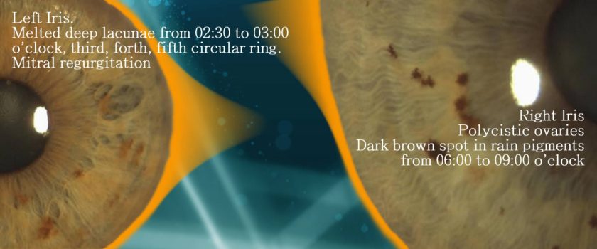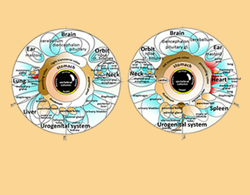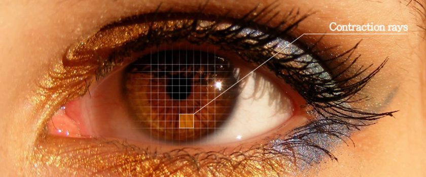
The anatomical sources on which iridology relies are three: the iris surface, the pupil and the sclera. Each of these structures provides information which integrates patient’s evaluation. Main iridology markings Lacunae. Oval or lozange shaped area without fibers or with very sparse one. Bright fibers. Lightening of texture. Transversal fibers. … Continue reading

THE BASICS OF IRIDOLOGY – A COLLECTION OF THREE HANDBOOKS ON IRIDOLOGY THE AUTHOR Francesco Puerari MD has worked in an Italian General Hospital’s Anesthesia and Intensive Care Unit for 34 years. He has earned several postgraduate specializations (Anesthesia and Intensive Care, Dietetics and Nutrition, Medical Toxicology, Neurology) at … Continue reading

IRIS RECOGNITION AND IRIDOLOGY Iris analysis is used in two main fields: Iris recognition and Iridology. Iris recognition is a biometric automated method used in the field of privacy and security. Like a snowflake, the iris is unique: a distinctive pattern that forms randomly in uterus in a process called chaotic … Continue reading

The iris is easy to examine. No invasive procedures are required, but simple ophthalmic instruments.
Iridology studies the colored portion of the eye named iris.
The iris is a highly innervated organ which is stimulated both by the external environment as well as by the body. Continue reading


